Home
Uncategories
Anatomy Of Chest Wall - Thoracic Wall And Breast Illustrations / Week chest wall (thoracic cage) anatomy component overview sternum manubrium body xiphoid process ribs to costal true ribs:
Anatomy Of Chest Wall - Thoracic Wall And Breast Illustrations / Week chest wall (thoracic cage) anatomy component overview sternum manubrium body xiphoid process ribs to costal true ribs:
Anatomy Of Chest Wall - Thoracic Wall And Breast Illustrations / Week chest wall (thoracic cage) anatomy component overview sternum manubrium body xiphoid process ribs to costal true ribs:. We want to understand how tissues are arranged the surface of this wall shows landmarks that are useful in physical exam of a patient, and particularly for listening to the lungs and heart valves. The embryologic and anatomic basis of modern surgery. Region in the trunk of the body that lies between the neck and… The layers of the chest wall include the skin, subcutaneous fat this chapter discusses the embryologic development and normal radiologic anatomy of the chest wall. The bony skeletal part of the thoracic wall is the rib cage, and the rest is made up of muscle, skin, and fasciae.
Various imaging techniques for evaluation of. Stability to arm and shoulder movement; And flexibility to aid in the functional process of respiration. A complete review of the left lateral chest. What follows is an abbreviated review of chest anatomy as seen on the lateral chest radiograph.
Chest Wall Lumps Rib Injury Clinic from www.ribinjuryclinic.com A working knowledge of their anatomy and of its variations is essential to any. Various imaging techniques for evaluation of. Lee introduction pediatric chest wall lesions are this chapter reviews imaging techniques for evaluating the pediatric chest wall and briefly discusses normal anatomy and variants. The chest anatomy includes the pectoralis major, pectoralis minor and the serratus anterior. Gest tr, chest wall anatomy. Stability to arm and shoulder movement; Week chest wall (thoracic cage) anatomy component overview sternum manubrium body xiphoid process ribs to costal true ribs: Understanding chest wall anatomy is paramount to any surgical procedure regarding the.
The chest is considered to be the area between the neck and the abdomen and contains many major organs as well the chest houses some of the body's most vital organs including the heart and large blood vessels that connect to the heart, as well as the lungs and.
Region in the trunk of the body that lies between the neck and… Documents similar to anatomy of the chest wall. Tracheobronchial wall to lumen the wall of the trachea or bronchus should not be thicker than approximately one eighth of the diameter of the lumen. A complete review of the left lateral chest. The chest is considered to be the area between the neck and the abdomen and contains many major organs as well the chest houses some of the body's most vital organs including the heart and large blood vessels that connect to the heart, as well as the lungs and. The chest wall is a complex system that provides rigid protection to the vital organs such as the heart, lungs, and liver; Chest wall anatomy (page 1). Bones of the thoracic wall. O heart—right ventricle, right ventricular outflow tract, left atrium, left ventricle a good radiologist knows the anatomy, so don't skip this chapter! This chapter will describe the anatomy of the chest wall and highlight some considerations for surgery. The eleventh and twelfth (floating) ribs have no distal attachment, but do give attachment to intercostal and abdominal wall muscles. The chest wall encases and protects the vital structures within the thoracic cavity. Principal functions are the protection of internal viscera and an expandable cylinder facilitating variable gas flow into the lungs.
Principal functions are the protection of internal viscera and an expandable cylinder facilitating variable gas flow into the lungs. How many organs could you technically live without? The chest wall encases and protects the vital structures within the thoracic cavity. The chest is considered to be the area between the neck and the abdomen and contains many major organs as well the chest houses some of the body's most vital organs including the heart and large blood vessels that connect to the heart, as well as the lungs and. Notice the expansile mass in the.
Anterior Thoracic Wall Anterior Thoracic Wall Internal View from netterimages.com Bones of the thoracic wall. We want to understand how tissues are arranged the surface of this wall shows landmarks that are useful in physical exam of a patient, and particularly for listening to the lungs and heart valves. Chest wall anatomy (page 1). This page provides an overview of the chest muscle group. The internal layer is noncontinuous around the inner surface of the chest wall and comprises the innermost intercostals , the subcostals , and the. Knowledge of the anatomy of the 'whole' cylinder (ribs, sternum, vertebra, diaphragm, intercostal spaces and extrathoracic muscles) is therefore not only important in the local environment of a specific chest wall resection but its relation to overall function. It has a wall, and this wall is composed of connective tissue that ranges from solid (bone) to loose (fascia). And flexibility to aid in the functional process of respiration.
The chest wall, like other regional anatomy, is a remarkable fusion of form and function.
This page provides an overview of the chest muscle group. Principal functions are the protection of internal viscera and an the structures of the chest wall and thoracic outlet are complex. Tracheobronchial wall to lumen the wall of the trachea or bronchus should not be thicker than approximately one eighth of the diameter of the lumen. And flexibility to aid in the functional process of respiration. Ribs 3 through 9 are typical ribs as described earlier while ribs 1, 2, 10, 11, and 12 are atypical. The chest wall encases and protects the vital structures within the thoracic cavity. Week chest wall (thoracic cage) anatomy component overview sternum manubrium body xiphoid process ribs to costal true ribs: Principal functions are the protection of internal viscera and an expandable cylinder facilitating variable gas flow into the lungs. The first rib is a short, flat rib that is much wider and more curved than those previously described. Region in the trunk of the body that lies between the neck and… O airway—trachea, upper lobe bronchi, posterior wall of bronchus intermedius. Documents similar to anatomy of the chest wall. Bones of the thoracic wall.
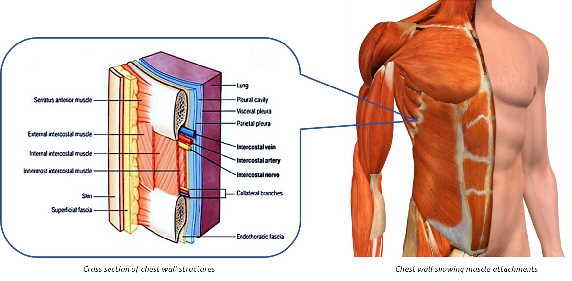
We want to understand how tissues are arranged the surface of this wall shows landmarks that are useful in physical exam of a patient, and particularly for listening to the lungs and heart valves. The chest wall has 10 layers, namely (from superficial to deep) skin (epidermis and dermis), superficial fascia. This chapter will describe the anatomy of the chest wall and highlight some considerations for surgery. Outward movements of chest wall. Paschalides medical publications, 2004, with permission.
Thoracic Wall And Breast Illustrations from www.imaios.com Surface anatomy of anterior chest wall. Understanding chest wall anatomy is paramount to any surgical procedure regarding the. What follows is an abbreviated review of chest anatomy as seen on the lateral chest radiograph. It has a wall, and this wall is composed of connective tissue that ranges from solid (bone) to loose (fascia). Gest tr, chest wall anatomy. Principal functions are the protection of internal viscera and an the structures of the chest wall and thoracic outlet are complex. Xiphoid process, costal arch, 12th and 11th ribs, vertebra t12. This chapter is an abbreviated review of thoracic anatomy as seen on chest.
Spiral ct of thoracic inlet.
Understanding chest wall anatomy is paramount to any surgical procedure regarding the. Region in the trunk of the body that lies between the neck and… Gest tr, chest wall anatomy. Various imaging techniques for evaluation of. Surface features & palpable landmarks o… 1. Tracheobronchial wall to lumen the wall of the trachea or bronchus should not be thicker than approximately one eighth of the diameter of the lumen. Paschalides medical publications, 2004, with permission. Stability to arm and shoulder movement; O airway—trachea, upper lobe bronchi, posterior wall of bronchus intermedius. Week chest wall (thoracic cage) anatomy component overview sternum manubrium body xiphoid process ribs to costal true ribs: The chest wall is a complex system that provides rigid protection to the vital organs such as the heart, lungs, and liver; The chest wall, like other regional anatomy, is a remarkable fusion of form and function. Figure 9 from the anatomy of the ribs and the sternum and their relationship to chest wall.
Ribs 3 through 9 are typical ribs as described earlier while ribs 1, 2, 10, 11, and 12 are atypical anatomy of chest. The bony skeletal part of the thoracic wall is the rib cage, and the rest is made up of muscle, skin, and fasciae.


0 Comments:
Posting Komentar